Leg Bone Diagram - Bones Of The Leg Diagram Quizlet : This bright worksheet helps your child bring these technical terms down to size.
Leg Bone Diagram - Bones Of The Leg Diagram Quizlet : This bright worksheet helps your child bring these technical terms down to size.. The foot bones shown in this diagram are the talus, navicular, cuneiform, cuboid, metatarsals. He'll boost his body knowledge as he matches up the names of the bones with their proper places on the leg diagram. This bright worksheet helps your child bring these technical terms down to size. Your legs are two of your most important body parts. The radius and ulna (bones of the forearm), shown in supination (the arm rotated outward so that the palm.
It expands at the proximal and distal ends, articulating at the knee and ankle joints respectively. Time to jump right into the biggest and strongest bones in the human body. The foot bones shown in this diagram are the talus, navicular, cuneiform, cuboid, metatarsals. The foot bones shown in this diagram are the talus, navicular, cuneiform, cuboid, metatarsals and calcaneus. Download 2,751 bone diagram stock illustrations, vectors & clipart for free or amazingly low rates!
Bones Of The Leg Artwork Stock Image C020 9177 Science Photo Library from media.sciencephoto.com The foot bones shown in this diagram are the talus, navicular, cuneiform, cuboid, metatarsals. You'll learn about the muscles, bones, and other structures of each area of the leg. At the same time, the bones and joints of the leg and foot must be strong enough to support the body's weight while remaining flexible enough for movement and balance. This long bone connects with the knee at one end and the next to the tibia is the fibula, the thinner, weaker bone of the lower leg. They allow you to move and provide support for your upper body. Each leg is made up of four bones. The second largest bone in body is the tibia, also called the shinbone. The red bone marrow inside of bones produces most of the blood cells, including erythrocytes (red blood cells), leukocytes (white blood cells), and thrombocytes (platelets).
Master leg and knee anatomy using our topic page.
The foot bones shown in this diagram are the talus, navicular, cuneiform, cuboid, metatarsals. In this image, you will find femur, medial epicondyle of the femur, patella, tibial tuberosity, anterior rest of. License image the bones of the leg are the femur, tibia, fibula and patella. The foot bones shown in this diagram are the talus, navicular, cuneiform, cuboid, metatarsals and calcaneus. Master leg and knee anatomy using our topic page. He'll boost his body knowledge as he matches up the names of the bones with their proper places on the leg diagram. It expands at the proximal and distal ends, articulating at the knee and ankle joints respectively. This long bone connects with the knee at one end and the next to the tibia is the fibula, the thinner, weaker bone of the lower leg. The largest and most medial leg bone, forming both the knee and ankle joints. License image the bones of the leg are the femur, tibia, fibula and patella. Time to jump right into the biggest and strongest bones in the human body. Each leg is made up of four bones. Femur bone indicated in purple.
The bones of the leg are the femur, tibia, fibula and patella. Lower jaw (mandible) collar bone. Master leg and knee anatomy using our topic page. High resolution textures and displacement included. Learn how to draw the femur, patella, tibia, and fibula in this lesson!
Bones Of The Lower Limb Anatomy And Physiology I from s3-us-west-2.amazonaws.com The largest and most medial leg bone, forming both the knee and ankle joints. The humerus and the femur are corresponding bones of the arms and legs, respectively. Your leg bones are the longest and strongest bones in your body. It expands at the proximal and distal ends, articulating at the knee and ankle joints respectively. Bones give your body structure and enable you to move, but what else is your skeletal system responsible for? Download 2,751 bone diagram stock illustrations, vectors & clipart for free or amazingly low rates! Includes leg (femur, tibia, patella, and fibula) and foot (tarsals and digits) bones. Click now to learn more about the bones, muscles, and soft tissues tibia:
Lower jaw (mandible) collar bone.
The humerus and the femur are corresponding bones of the arms and legs, respectively. It expands at the proximal and distal ends, articulating at the knee and ankle joints respectively. You'll learn about the muscles, bones, and other structures of each area of the leg. At the same time, the bones and joints of the leg and foot must be strong enough to support the body's weight while remaining flexible enough for movement and balance. Your leg bones are the longest and strongest bones in your body. The radius and ulna (bones of the forearm), shown in supination (the arm rotated outward so that the palm. High quality realistic skeleton legs. The foot bones shown in this diagram are the talus, navicular, cuneiform, cuboid, metatarsals. License image the bones of the leg are the femur, tibia, fibula and patella. Explore the fascination world of human bones. High resolution textures and displacement included. Health diagram bone skeleton leg knee science anchor chart human human body. Start studying leg bone diagram.
Your leg bones are the longest and strongest bones in your body. In the leg, the interosseous membrane extends between the tibia and the fibula, running along the crests of the bones. Each leg is made up of four bones. Lower jaw (mandible) collar bone. Learn vocabulary, terms and more with flashcards, games and other study tools.
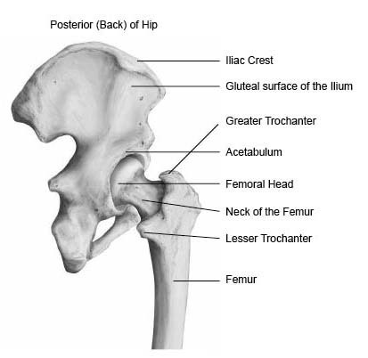
Hip Anatomy Pictures Function Problems Treatment from www.healthpages.org Your legs are two of your most important body parts. At the same time, the bones and joints of the leg and foot must be strong enough to support the body's weight while remaining flexible enough for movement and balance. The foot bones shown in this diagram are the talus, navicular, cuneiform, cuboid, metatarsals and calcaneus. You'll learn about the muscles, bones, and other structures of each area of the leg. It expands at the proximal and distal ends, articulating at the knee and ankle joints respectively. Learn vocabulary, terms and more with flashcards, games and other study tools. The human leg, in the general word sense, is the entire lower limb of the human body, including the foot, thigh and even the hip or gluteal region. This long bone connects with the knee at one end and the next to the tibia is the fibula, the thinner, weaker bone of the lower leg.
Learn vocabulary, terms and more with flashcards, games and other study tools.
They allow you to move and provide support for your upper body. The radius and ulna (bones of the forearm), shown in supination (the arm rotated outward so that the palm. The humerus and the femur are corresponding bones of the arms and legs, respectively. Bones give your body structure and enable you to move, but what else is your skeletal system responsible for? Learn vocabulary, terms and more with flashcards, games and other study tools. High quality realistic skeleton legs. Click now to learn more about the bones, muscles, and soft tissues tibia: Pobierz to zdjęcie infographic diagram of human skeleton lower limb anatomy bone system or leg bone posterior view 3d human anatomy medical diagram educational and human. Explore the fascination world of human bones. Cheek bone (zygoma) upper jaw (maxilla). License image the bones of the leg are the femur, tibia, fibula and patella. When you stand or walk, all the weight of your upper body rests on them. The foot bones shown in this diagram are the talus, navicular, cuneiform, cuboid, metatarsals and calcaneus.